Application of infrared thermography for animal health study
DOI:
https://doi.org/10.31783/elsr.2022.81152157Keywords:
animal, health, infrared thermography, stress, temperatureAbstract
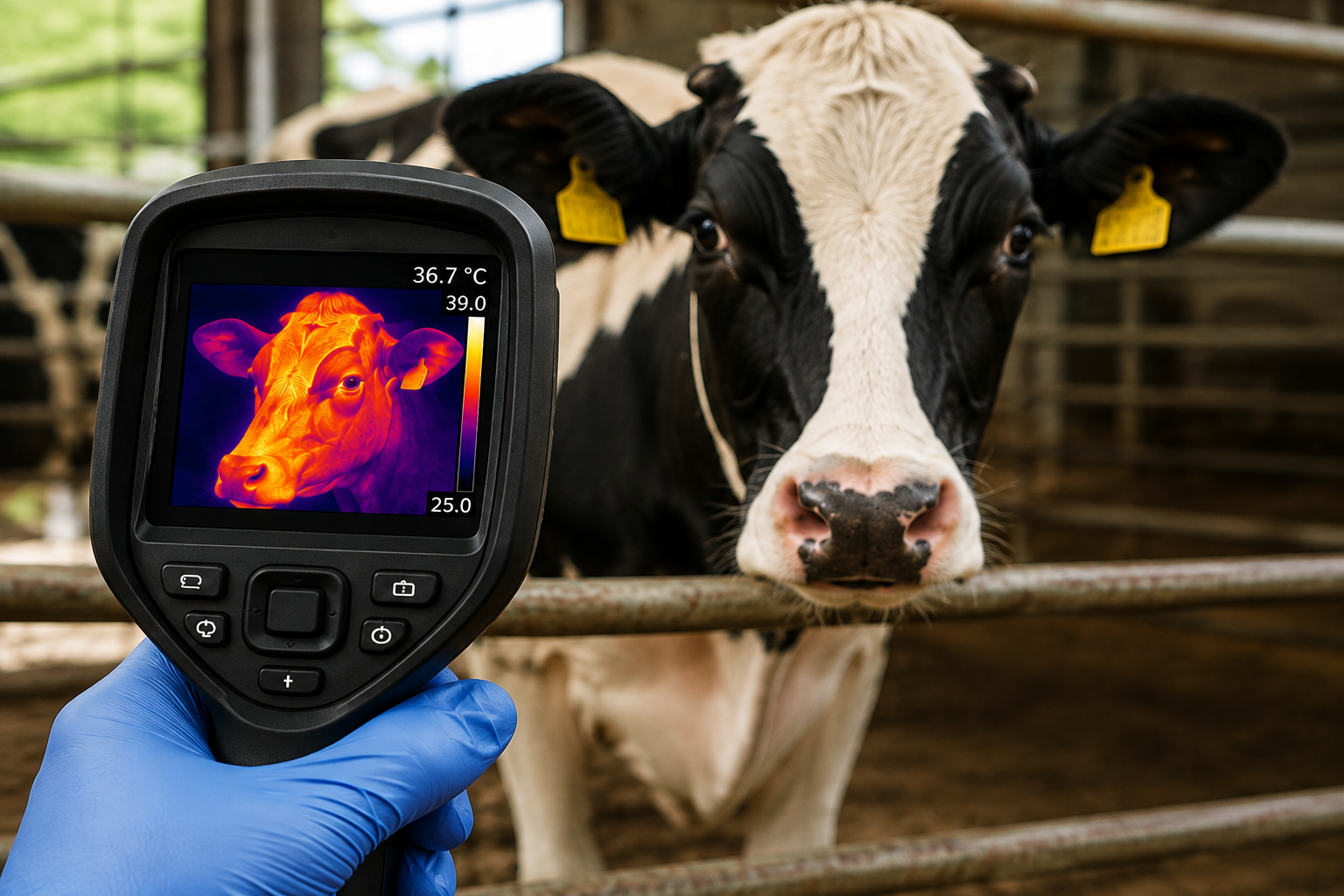
Infrared thermography (IRT) is an advanced, safe, non-invasive, and contactless technology used to determine the surface temperature of an object. Thermal cameras absorb the infrared radiation generated by the surface, then transform it into electrical signals, and provide a thermal image of the body's surface temperature distribution. In addition to human medicine, IRT has mostly been used in veterinary medicine for diagnostic purposes and to detect distress in an animal. However, IRT can be a very effective technique for livestock and poultry breeding research viz. thermoregulation, reproduction, animal welfare, etc. Before pre-clinical testing, it is crucial to intervene to reduce the stress levels in experimental animals for the betterment of the experimental outcome. Where the infrared thermography could help us to monitor and assess the health status or the stress level in addition to behavioral and physiological parameters before the pre-clinical testing might be beneficial for better outcome and reproducibility. Therefore, this manuscript reviews the recent advances in infrared thermography and its use in animal science.
References
[1]. S. K. Yadav, P. Singh, P. Kumar, S. V. Singh, A. Singh and S. Kumar (2019). Scrotal infrared thermography and testicular biometry: Indicator of semen quality in murrah buffalo bulls. Anim. Reprod. Sci., 209: 106145. doi: 10.1016/j.anireprosci.2019.106145.
[2]. F. A. Barca Junior, C. K. Junior, P. da Cruz Fávaro, G. R. Pereira, S. R. O. Menegassi, F. Morotti and V. H. G. Galdioli et al., (2020). Infrared thermography and Doppler ultrasonography to evaluate the effects of scrotal insulation on testicular blood flow dynamics in bulls. Semina: Cienc. Agrar., 41: 1267-1278.
[3]. K. A. Herborn, J. L. Graves, P. Jerem, N. P. Evans, R. Nager, D. J. McCafferty and D. E. F. McKeegan (2015). Skin temperature reveals the intensity of acute stress. Physiol. Behav., 152: 225-230.
[4]. P. Carrive, M. Churyukanov and D. Le Bars (2011). A reassessment of stress-induced “analgesia” in the rat using an unbiased method. Pain, 152: 676-686.
[5]. F. Kano, S. Hirata, T. Deschner, V. Behringer and J. Call (2015). Nasal temperature drop in response to a playback of conspecific fights in chimpanzees: A thermo-imaging study. Physiol. Behav., 155: 83-94.
[6]. J. Weitkamp (2020). Effect of tickling and gentling on eye and tail temperature of laboratory rats during manual restraint, using infrared thermography. Master’s Thesis, Utrecht University, Utrecht, The Netherlands, March 2020.
[7]. N. Ludwig, M. Gargano, F. Luzi, C. Carenzi and M. Verga (2007). Technical note: Applicability of infrared thermography as a non invasive measurements of stress in rabbit. World Rabbit Sci., 15: 199-206.
[8]. K. Fiebig, T. Jourdan, M. H. Kock, R. Merle and C. Thöne-Reineke (2018). Evaluation of infrared thermography for temperature measurement in adult male NMRI nude mice. J. Am. Assoc. Lab. Anim. Sci., 57: 715-724.
[9]. F. R. De Ruediger, P. H. Yamada, L. G. B. Barbosa, M. G. M. Chacur, J. C. P. Ferreira, N. A. T. de Carvalho and G. A. M. Soriano et al., (2018). Effect of estrous cycle phase on vulvar, orbital area and muzzle surface temperatures as determined using digital infrared thermography in buffalo. Anim. Reprod. Sci., 197: 154-161.
[10]. S. C. Scolari, S. G. Clark, R. V. Knox and M. A. Tamassia (2011). Vulvar skin temperature changes significantly during estrus in swine as determined by digital infrared thermography. J. Swine Health Prod., 19: 151-155.
[11]. A. L. Schaefer, N. J. Cook, C. Bench, J. B. Chabot, J. Colyn, T. Liu and E. K. Okine et al., (2012). The non-invasive and automated detection of bovine respiratory disease onset in receiver calves using infrared thermography. Res. Vet. Sci., 93: 928-935.
[12]. E. Bartolomé, F. Azcona, M. Cañete-Aranda, D.I. Perdomo-González, J. Ribes-Pons and E.M. Terán (2019). Testing eye temperature assessed with infrared thermography to evaluate stress in meat goats raised in a semi-intensive farming system: a pilot study. Arch. Anim. Breed., 62: 199-204.
[13]. K. L. Farrar, A. E. Field, S. L. Norris and K. O. Jacobsen (2020). Comparison of rectal and infrared thermometry temperatures in anesthetized swine (Sus scrofa). J. Am. Assoc. Lab. Anim. Sci., 59: 221-225.
[14]. R. D. S. Sousa, F. L. C. de Oliveira, M. R. B. Dias, N. S. Minami, L. D. Amaral, A. H. H. Minervino and C. D. L. Shecaira (2020). Evaluation of infrared thermography, force platform and filmed locomotion score as non-invasive diagnostic methods for acute laminitis in zebu cattle. Plos One, 15: e0235549. doi: 10.1371/journal.pone.0235549.
[15]. A. Yáñez-Pizaña, D. Mota-Rojas, R. Ramírez-Necoechea, M. Castillo-Rivera, P. Roldán-Santiago, P. Mora-Medina and M. González-Lozano (2019). Application of infrared thermography to assess the effect of different types of environmental enrichment on the ocular, auricular pavilion and nose area temperatures of weaned piglets. Comput. Electron. Agric., 156: 33-42.
[16]. L. M. Rocha, N. Devillers, X. Maldague, F. Z. Kabemba, J. Fleuret, F. Guay and L. Faucitano (2019). Validation of anatomical sites for the measurement of infrared body surface temperature variation in response to handling and transport. Animals (Basel), 9: 425. doi:10.3390/ani9070425.
[17]. A. Colak, B. Polat, Z. Okumus, M. Kaya, L. E. Yanmaz and A. Hayirli (2008). Early detection of mastitis using infrared thermography in dairy cows. J. Dairy Sci., 91: 4244-4248.
[18]. B. Yadav, G. Singh and A. Wankar (2017). The use of infrared skin temperature measurements for monitoring heat stress and welfare of crossbred cattle. Indian J. Dairy Sci., 70: 127-131.
Downloads
Published
Issue
Section
License
Copyright (c) 2022 Author (s)

This work is licensed under a Creative Commons Attribution-NonCommercial-NoDerivatives 4.0 International License.
Copyright © The Author(s). This is an open access article distributed under the terms of the Creative Commons Attribution-NonCommercial-NoDerivatives 4.0 International License (CC BY-NC-ND 4.0), which permits unrestricted non-commercial use, distribution, and reproduction in any medium, provided the original author(s) and source are properly credited, and the work is not modified or adapted.