Stability analysis of nano-dispersed food bio-colour extracted from coconut milk as a substrate by using yeast (Xanthophyllomyces dendrorus)
DOI:
https://doi.org/10.31783/elsr.2023.910109Keywords:
astaxanthin, biocolour, carotenoids, xanthophyllomyces dendrorusAbstract
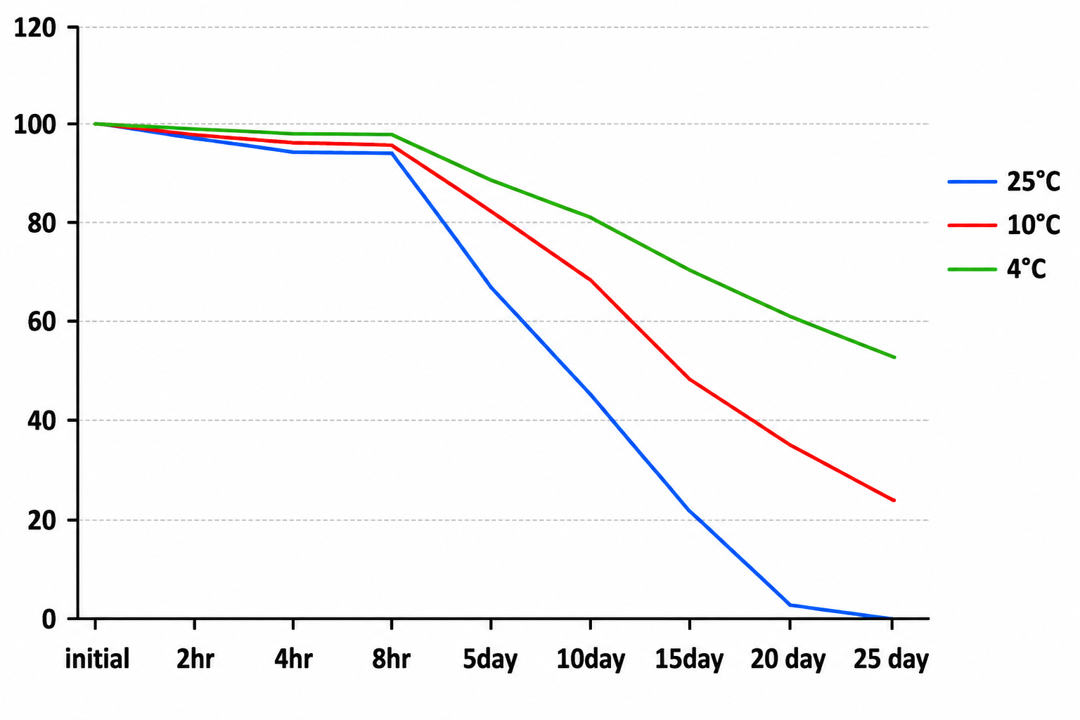
Food color plays a prominent role in the sensory evaluation of any food materials; because of this it affects the uniqueness, desirability, and palatability of food. Properties such as easy synthesis and cost-effectiveness made synthetic colors used more in different kinds of food. Many synthetic dyes have been causing several adverse effects and toxic diseases including Attention Deficit Hyperactivity Disorder (ADHD), hyper-allergenicity, food allergies, depression, headaches, carcinogenicity, migraines, and other toxicological issues like issues related to children's behavior. This has been a driving force for many food technologists all over the world to show interest in replacing synthetic colors with natural colorants as per the market demand as well as clean-label product promotion. Biocolors are not only color foods but also have medical and therapeutic effects. They are derived from natural sources including plants, animals, algae, fungi, and insects that can impart certain colors to foods. Microorganisms are classified as a highly significant source for biocolour production due to their merits like durability, availability, labor, cost-effectiveness, productivity, and simple downstream processing in comparison with plants. Coconut milk was used as the substrate for biocolour preparation by using yeast(Xanthophyllomyces dendrorus), where maximum biomass yield was 5.15±0.096 g/L, extracted colour (astaxanthin) yield of 0.84±0.024 g/L and maximum nano-dispersed color yield 0.83±0.024g shows efficiency more than 90% was produced. The Nano-dispersed color incubated under conditions including varied light intensities or conditions, different pH, and different temperatures were observed with degradation of biocolour with p<0.01 high significance.
References
[1] S. Ghosh, T. Sarkar, A. Das and R. Chakraborty (2022). Natural colorants from plant pigments and their encapsulation: An emerging window for the food industry. LWT - Food Sci. Technol., 153: 112527. doi: 10.1016/J.LWT.2021.112527.
[2] S. Ganguly, K. Bandyopadhyay, P. Dutta and A. Pramanik (2019). A review on microbial-pigment: A good source of biocolour. Pharma Innov. J., 8: 416-420.
[3] P. Bora, P. Das, R. Bhattacharyya and M. S. Barooah (2019). Biocolour: The natural way of colouring food. J. Pharmacogn. Phytochem., 8: 3663-3668.
[4] C. Gupta, A. P. Garg, D. Prakash, S. Goyal and S. Gupta (2011). Microbes as potential source of biocolours. Pharmacology., 2: 1309-1318.
[5] H. S. Tuli, P. Chaudhary, V. Beniwal and A. K. Sharma (2015). Microbial pigments as natural color sources: current trends and future perspectives. J. Food Sci. Technol., 52: 4669-4678.
[6] S. Samyuktha and S. N. Mahajan (2016). Isolation and identification of pigment producing bacteria and characterization of extracted pigments. Int. J. Appl. Res., 2: 657-664
[7] Z.-C. Hu, Y. G. Zheng, Z. Wang and Y. C. Shen (2007). Production of astaxanthin by Xanthophyllomyces dendrorhous ZJUT46 with fed-batch fermentation in 2.0 M3 fermentor. Food Technol. Biotechnol., 45: 209-212.
[8] V. Monishdeep, H. W. Deshpande, A. Poshadri and B. V. P. Kumar (2022). A review on potential microbial sourced bio-colours from fungi. Pharma Innov. J., 11(Special Issue): 3200-3205.
[9] N. F. Haard (1988). Astaxanthin formation by the yeast Phaffia rhodozyma on molasses, Biotechnology Lett., 10: 609-614.
[10] J. D. Fontana, M. F. Guimaraes, N. T. Martins, C. A. Fontana and M. Baron (1996). Culture of the astaxanthinogenic yeast phaffia rhodozyma in low-cost media. Appl. Biochem. Biotechnol., 57-58: 413-422.
[11] J. D. Fontana, D. A. Mitchell, O. E. Molina, A. Gaitan, T. M. B. Bonfim, J. Adelmann and A. Grzybowski (2008). Starch depolymerization with diluted phosphoric acid and application of the hydrolysate in Astaxanthin fermentation. Food Technol. Biotechnol., 46: 305-310.
[12] M. Vázquez, F. Diniz and A. M. Martin (1996). Single-cell protein from Xanthophyllomyces dendrorhous yeast: continuous fermentation using peat extract as substrate. Ciencia Tecnol. Alime., 2: 12-16.
[13] M. Olaizola (2000). Commercial production of astaxanthin from Haematococcus pluvialis using 25,000-liter outdoor photobioreactors. J. Appl. Phycol., 12: 499-506.
[14] J. Ramırez, M. L. Nunez and R. Valdivia (2000). Increased astaxanthin production by a Phaffia rhodozyma mutant grown on date juice from Yucca fillitera. J. Ind. Microbiol. Biotechnol., 24: 187-190.
[15] C. H. Luna-Flores, J. J. Ramrrez-Cordova, C. Pelayo-Ortiz and E. J. HerreraLopez (2010). Batch and fed-batch modeling of carotenoids production by Xanthophyllomyces dendrorhous using Yucca fillifera date juice as substrate. Biochem. Eng. J., 53: 131-136.
[16] A. R. Dominguez-Bocanegra and J. A. Torres-Muñoz (2005). Astaxanthin hyperproduction by Phaffia rhodozyma (now Xanthophyllomyces dendrorhous) with raw coconut milk as sole source of energy. Appl. Microbiol. Biotechnol., 66: 249-252.
[17] R. J. Stoklosa, D. B. Johnston and N. P. Nghiem (2019). Phaffia rhodozyma cultivation on structural and non-structural sugars from sweet sorghum for astaxanthin generation. Process Biochem., 83: 9-17.
[18] A. Torres-Haro, A. Gschaedler, J. C. Mateos-Díaz, E. J. Herrera-López, R. M. Camacho-Ruíz and M. Arellano-Plaza (2021). Improvement of a specific culture medium based on industrial glucose for carotenoid production by Xanthophyllomyces dendrorhous. Processes, 9: 429. doi: 10.3390/pr9030429.
[19] K. Jirasripongpun, W. Pewiong, P. Kitraksa and C. Krudngern (2008). Carotenoid production by Xanthophyllomyces dendrorhous: use of pineapple juice as a production medium, Lett. Appl. Microbiol., 47: 112-116.
[20] N. Ananda and P. V. Vadlani (2010). Production and optimization of carotenoid-enriched dried distiller’s grains with solubles by Phaffia rhodozyma and Sporobolomyces roseus fermentation of whole stillage. J. Ind. Microbiol. Biotechnol., 37: 1183-1192.
[21] B. Stachowiak (2012). Astaxanthin synthesis by yeast Xanthophyllomyces dendrorhous and its mutants on media based on plant extracts. Indian J. Microbiol., 52: 654-659.
[22] C. Yuan, L. Du, Z. Y. Jin, X. M. Xu Storage stability and antioxidant activity of complex of astaxanthin with hydroxypropyl-β-cyclodextrin, Carbohydr. Polym., 91: 385-389.
[23] Q. Shen and S. Y. Quek (2014). Microencapsulation of astaxanthin with blends of milk protein and fiber by spray drying. J. Food Eng., 123: 165-171.
[24] P. C. Mezquita, B. E. B. Huerta, J. C. P. Ramírez and C. P. O. Hinojosa (2015). Milks pigmentation with astaxanthin and determination of colour stability during short period cold storage. J. Food Sci. Technol., 52: 1634-1641.
[25] F. Tamjidi, M. Shahedi, J. Varshosaz and A. Nasirpour (2018). Stability of astaxanthin-loaded nanostructured lipid carriers in beverage systems. J. Sci. Food Agric., 98: 511-518.
[26] M. Vakarelova, F. Zanoni, P. Lardo, G. Rossin, F. Mainente, R. Chignola and A. Menin et al., (2016). Production of stable food-grade microencapsulated astaxanthin by vibrating nozzle technology. Food Chem., 221: 289-295.
[27] A. Bassijeh, S. Ansari and S. M. H. Hosseini (2020). Astaxanthin encapsulation in multilayer emulsions 1283 stabilized by complex coacervates of whey protein isolate and Persian gum and its use as a 1284 natural colorant in a model beverage. Food Res. Int., 137: 109689. doi: 10.1016/j.foodres.2020.109689.
[28] N. Anarjan and C. P. Tan (2013). Chemical stability of astaxanthin nanodispersions in orange juice and skimmed milk as model food systems. Food Chem., 139: 527-531.
[29] N. Anarjan and C. P. Tan (2013). Effects of storage temperature, atmosphere and light on chemical stability of astaxanthin nanodispersions, J. Am. Oil Chem. Soc., 90: 1223-1227.
[30] S. R. Mhalaskar, S. S. Thorat and A. A. Kulthe (2018). Stability analysis of food bio-colour extracts from broken rice through solid state fermentation. Int. J. Pure App. Biosci., 6: 304-313.
Downloads
Published
Issue
Section
License
Copyright (c) 2023 Author (s)

This work is licensed under a Creative Commons Attribution-NonCommercial-NoDerivatives 4.0 International License.
Copyright © The Author(s). This is an open access article distributed under the terms of the Creative Commons Attribution-NonCommercial-NoDerivatives 4.0 International License (CC BY-NC-ND 4.0), which permits unrestricted non-commercial use, distribution, and reproduction in any medium, provided the original author(s) and source are properly credited, and the work is not modified or adapted.