Pre- and post- morphological evaluation of the kidneys of male Wistar rats inoculated with Rhipicephalus sanguineus (Latreille, 1806) (Acari: Ixodidade) salivary gland extract
DOI:
https://doi.org/10.31783/elsr.2018.421121Keywords:
bioactive, control, pathogen, R. sanguineus, salivary gland, ticksAbstract
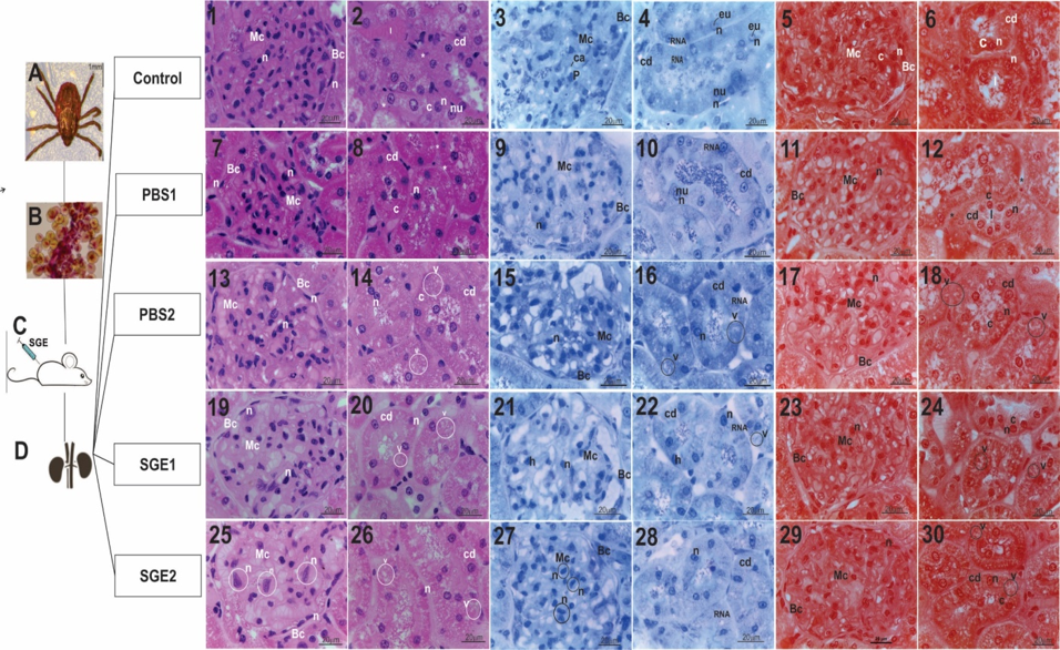
Controlling the population of ticks is a subject of great concern to researchers, due to the parasitic nature of these ectoparasites and their capacity to transmit pathogens to the human being and other vertebrate animals. In this scenario, some authors have studied the salivary glands of Rhipicephalus sanguineus, having demonstrated the morphophysiological importance of these organs. The saliva plays a role in the modulation of the host’s immune-inflammatory and hemostatic systems; in addition, it is considered a potential reservoir of several functions. In this sense, the present study had the objective to establish, analyze and correlate the morpho-physiological behavior of the cells and tissues from the kidneys of male Wistar rats pre/post inoculated with R. sanguineus salivary gland extract, in addition to verify the possible toxic action of this saliva on the kidney. For this, the rats were divided into 5 groups with 4 animals each: CG (control, no exposure); PBS1 (one injection of PBS 1X), PBS2 (two injections of PBS 1X), SGE1 (one injection of salivary gland extract 0.04µg/µL) and SGE2 (two injections of salivary gland extract 0.04µg/µL). After the exposures, the kidneys were subjected to histological/histochemical techniques: HE, toluidine blue and xylidine Ponceau. Each inoculated rat was weighed one day before the first inoculation and after 21 days, the blood count analysis was performed.
References
[1] W. R. Kaufman (1989). Tick-host interaction: a synthesis of current concepts. Parasitol. Today, 5: 47-56.
[2] D. E. Sonenshine (1991). The female reproductive system. In: D. E. Sonenshine (Org.). Biology of Ticks. New York: Oxford University Press, pp. 280-304.
[3] F. Dantas-Torres (2010). Biology and ecology of the brown dog tick, Rhipicephalus sanguineus. Parasit Vectors, 3: 26. doi.org/10.1186/1756-3305-3-26.
[4] Y. S. Balashov (1967). Blood-sucking ticks (Ixodoidea)-vectors of diseases of man and animals. Blood-sucking ticks (Ixodoidea)-vectors of diseases of man and animals.
[5] M. I. Camargo-Mathias (2013). Guia básico de morfologia interna de carrapatos ixodídeos. São Paulo: Editora Unesp, 120 pp.
[6] K. C. S. Furquim (2007). Estudo das glândulas salivares de fêmeas e de machos de carrapatos Rhipicephalus sanguineus (Latreille, 1806) (Acari, Ixodidae): caracterização do ciclo secretor com ênfase no processo de degeneração. Tese de Doutorado, Instituto de Biociências, UNESP, Rio Claro, Brazil.
[7] M. I. Camargo-Mathias and K. C. S. Furquim (2013). The histology as a tool for the understanding of the morphophysiology of the brown dog tick (Rhipicephalus sanguineus). In: Jenkins, O.P (Ed), Advances in Zoology Research. USA: Nova Science Publishers Inc., 8: 167-191.
[8] K. C. S. Furquim, G. H. Bechara and M. I. Camargo-Mathias (2008a). Degeneration of salivary glands of males of the tick Rhipicephalus sanguineus (Latreille, 1806) (Acari, Ixodidae). Vet. Parasitol., 154: 325-335.
[9] K. C. S. Furquim, G. H. Bechara and M. I. Camargo-Mathias (2008b). Markers of cell death in salivary glands of males of the tick Rhipicephalus sanguineus (Latreille, 1806) (Acari, Ixodidae). Parasitol. Int., 57: 396-404.
[10] K. C. S. Furquim, G. H. Bechara and M. I. Camargo-Mathias (2010). Morpho-histochemical caracterization of salivar gland cells of males of the tick Rhipicephalus sanguineus (Acari, Ixodidae) at different feeding stages: description of new cell types. Exp. Appl. Acarol., 50: 59-70.
[11] B. S. Feldman-Muhsam and S. Saliternik-Givante (1970). Salivary secretion of the male tick during copulation. J. Insect Physiol., 16: 1945-1949.
[12] M. I. Camargo-Mathias, K. C. S. Furquim and P. H. Nunes (2011). Immunomodulatory effects of tick saliva. Invertebrate Survival Journal, 8: 231-240.
[13] N. A. Steen, S. C. Barker and P. F. Alewood (2006). Proteins in the saliva of the Ixodida (ticks): pharmacological features and biological significance. Toxicon, 47: 1-20.
[14] I. M. Francischetti, A. Sá-Nunes, B. J. Mans, L. M. Santos and J. M. Ribeiro (2010). The role of saliva in tick feeding. Frontiers in Bioscience: a journal and virtual library, 14: 2051-2088.
[15] C. J. F. Oliveira, W. A. Carvalho, G. R. Garcia, F. R. Gutierrez, I. K. de Miranda Santos, J. S. Silva and B. R. Ferreira (2010). Tick saliva induces regulatory dendritic cells: MAP-kinases and Toll-like receptor-2 expression as potential targets. Vet. Parasitol., 167: 288-297.
[16] M. Kazimirová and I. Stibraniová (2013). Tick salivary compounds: their role in modulation host defenses and pathogen transmission. Front. Cell. Infect. Microbiol., 3: 43. doi: 10.3389/fcimb.2013.00043.
[17] A. C. P. Volp, I. R. T. Renhe, P. C. Stringueta (2009). Pigmentos Naturais Bioativos, Ali. Nutr. Araraquara, Brazil, 20: 157-166.
[18] M. Kazimirová, I. Dovinová, T. Rolniková, L. Tóthová and L. Hunáková (2006). Anti-proliferative activity and apoptotic effect of tick salivary gland extracts on human HeLa cells. Neuro. endocriminol. Lett., 27 Suppl., 2: 48-52.
[19] J. R. Sauer, J. L. Mcswain, A. S. Bowman and R. C. Essenberg (1995). Tick salivary gland phyology. Annu. Rev. Entomol., 40: 245-267.
[20] E. Behling-Kelly and C. J. Czuprynski (2007). Endothelial cell as active participants in veterinary infections and inflammatory disorders. Anim. Health Res. Rev., 8: 47-58.
[21] S. K. Wikel and J. R. Allen (1977). Acquired resistance to ticks. III. Cobra venom factor and the resistance response, Immunology, 32: 457-465.
[22] L. C. Junqueira and J. Carneiro (2013). Histologia Básica – Texto & Atlas. 12ª Ed. São Paulo: Editora Guanabara Koogan, 528pp.
[23] M. R. Abreu, F. A. Rocha, K. C. S. Furquim, L. A. Anholeto, F. C. F. Novaes, M. J. Morsoleto and M. I. Camargo-Mathias (2014). Salivary Glands of Female Ticks Rhipicephalus sanguineus Like a Potential Source of Molecules with Inhibitory Action: In vivo study with Walker 256 Tumor Cells. Journal of Pharmaceutical Care & Health Systems, 1, 1-8. doi: 10.4172/2376-0419.1000121.
[24] J. Sedmak and S. E. Grossberg (1977). A rapid sensitive and versatile assay for protein using Coomassie Brilhant Blue G250. Anal. Biochem., 79: 544-552.
[25] L. C. U. Junqueira and L. M. M. S. Junqueira (1983). Técnicas básicas de citologia e histologia. São Paulo: Santos. Editora Santos, pp 123.
[26] M. L. S. Mello and B. C. Vidal (1980). Práticas de Biologia Celular. Edgard Blucher, São Paulo, Brazil.
[27] B. C. Vidal and M. L. S. Mello (1987). Biologia Celular. São Paulo, Brazil. Ed. Atheneu, pp 347.
[28] M. L. S. Mello, B. C. Vidal, M. M. Dantas and A. L. P. Monteiro (1993). Discrimination of the nucleolus by a Critycal Electrolyte Concentration method. Acta Histochem Cytochem, 26: 1-3.
[29] L. Lison (1960). Histochimie et cytochimie animales – Principles et méthodes, Gauthier Villars, Paris, 1.
[30] M. L. S. Mello and B. C. Vidal (1973). Linear dichoism and anomalus dispersions of birefringence on sperm heads. Acta Histochemica, 45: 109-114.
[31] M. L. S. Mello (1987). Nuclear cytochemistry and polarization microscopy of the spermatozoa of Triatoma infestans Klug. Z. Mikrosk. Anat. Forsch Leipzig, 101, 245-250.
[32] OECD (2001). Guideline 423: Acute Oral Toxicity - Acute Toxic Class Method. Acessado em 06 de janeiro de 2016 em http:// http://www.keepeek.com/Digital-Asset-Management/oecd/environment/test-no-423-acute-oral-toxicity-acute-toxic-class-method_9789264071001-en#page1 Paris: Head of Publications Service. doi : 10.1787/9789264071001-en.
[33] F. H. D. González and S. C. SILVA, S. C (2003). Introdução à Bioquímica Clínica Veterinária. Porto Alegre: UFRGS, 179-198.
Downloads
Published
Issue
Section
License
Copyright (c) 2018 Author (s)

This work is licensed under a Creative Commons Attribution-NonCommercial-NoDerivatives 4.0 International License.
Copyright © The Author(s). This is an open access article distributed under the terms of the Creative Commons Attribution-NonCommercial-NoDerivatives 4.0 International License (CC BY-NC-ND 4.0), which permits unrestricted non-commercial use, distribution, and reproduction in any medium, provided the original author(s) and source are properly credited, and the work is not modified or adapted.